
Why I use ultrasound to guide my injections — and what it means for you
What ultrasound shows us beneath the skin, why anatomy varies between individuals, and how that information supports more careful, considered treatment planning.
TL;DR Facial anatomy varies from person to person. Blood vessels, nerves, tissue layers, and previous filler do not always sit where textbooks suggest they should. Ultrasound allows me to assess your individual anatomy in real time and use that information to guide more careful, considered treatment planning. I have used ultrasound throughout my career as an anaesthetist, and I bring that same precision-focused approach to aesthetic medicine.
Patients often assume that facial anatomy is predictable — that vessels, nerves, and tissue planes sit in the same place for everyone. In reality, every face is different. That is one of the reasons I use ultrasound in my aesthetic practice: it allows me to assess the anatomy in front of me, rather than relying on textbook averages alone.
Other injectors — nurses and doctors alike — are often curious about this approach, because ultrasound is still uncommon in cosmetic practice. For me, it is a natural extension of how I have worked for years as an anaesthetist.
Why it matters more than most practitioners let on:
What we’re working with
The face is not a simple structure. Beneath the skin sits a layered architecture: fat compartments that shift with age, muscles that express emotion and chew and squint, ligaments that anchor skin to bone, and — running through all of it — an intricate network of blood vessels and nerves.
Careful treatment planning depends on understanding where a needle tip sits in relation to vessels, nerves, tissue planes, and previous filler.
Historically, practitioners have navigated this space using anatomical knowledge alone — memorising the expected locations of structures based on cadaver studies and textbooks. That knowledge is essential. I rely on it every day.
But here is the thing about human faces: they are not textbooks.
2. Why anatomy varies — and why that matters
Every patient who comes to see me has a face shaped by their genetics, their age, their sun exposure, prior treatments, and a thousand other individual factors. The angular artery that typically runs in a particular location may, in a given individual, take a course several millimetres from where a textbook would place it. Fat compartments shrink and shift. Filler placed in previous treatments occupies space and changes tissue planes.
I have a background in regional anaesthesia — a discipline where I spend a significant portion of my working life placing needles precisely near nerves, vessels, and vital structures using real-time ultrasound guidance. The philosophy is the same: when you can see what you are doing, you make better decisions.
Ultrasound lets me visualise soft tissue layers in real time, before and during treatment. That means I can identify where vessels lie in this patient, today — not where they are supposed to be on average. I can see how tissue moves with needle advancement. I can confirm filler placement within the intended plane. And if something looks unexpected, I can pause and think before proceeding.
3. What ultrasound actually shows
A modern portable ultrasound probe placed on the face reveals considerably more than most patients imagine.
The skin layers — epidermis, dermis, subcutaneous fat — are visible as distinct bands. Deeper fat compartments and the fascial layers that separate them can be differentiated. Muscles appear as characteristic striated structures. Blood vessels — arteries and veins — are identifiable by their appearance and, with Doppler function, by the blood flow within them.
HA filler, whether freshly placed or from previous treatments, is visible as a hypoechoic (darker) material within the tissue. This is significant: I can see not only where I am placing product, but what is already there from treatments performed elsewhere.
Nerves may be visible, particularly the larger named branches. Bony landmarks and ligaments can be identified. The whole exercise of treatment planning becomes less a matter of memory and more a matter of direct observation.
4. This is not routine practice — yet
I want to be honest with you: most cosmetic injectors do not use ultrasound. The technology requires training to interpret, takes time to integrate into a consultation, and demands investment in equipment. Many practitioners produce excellent results without it, particularly in lower-risk treatment areas.
But serious complications in aesthetic medicine — vascular occlusion, tissue necrosis, visual disturbance — are not hypothetical. They are documented, they are reported, and they are more likely in anatomically complex areas or in patients with prior filler. The literature on ultrasound guidance in aesthetics is growing, and several of the field’s most experienced practitioners argue it represents the direction the specialty should move.
I have watched this shift happen before. When I trained in anaesthesia, ultrasound guidance for procedures like central line insertion and nerve blocks was far from universal. Over the course of my career, it has become the expected standard — because the evidence and clinical practice evolved, and the profession moved towards direct visualisation where appropriate. Central lines are now routinely placed under direct ultrasound vision. Peripheral nerve blocks for surgery are guided in real time. The idea of doing these procedures blind, when you don’t have to, has become difficult to justify.
I expect the same trajectory in aesthetics. The evidence base is building. The technology is accessible. And as more practitioners with imaging backgrounds enter the field, the question will shift from why would you use ultrasound? to why wouldn’t you?
We are not there yet. But I would rather be ahead of that curve than catch up to it.
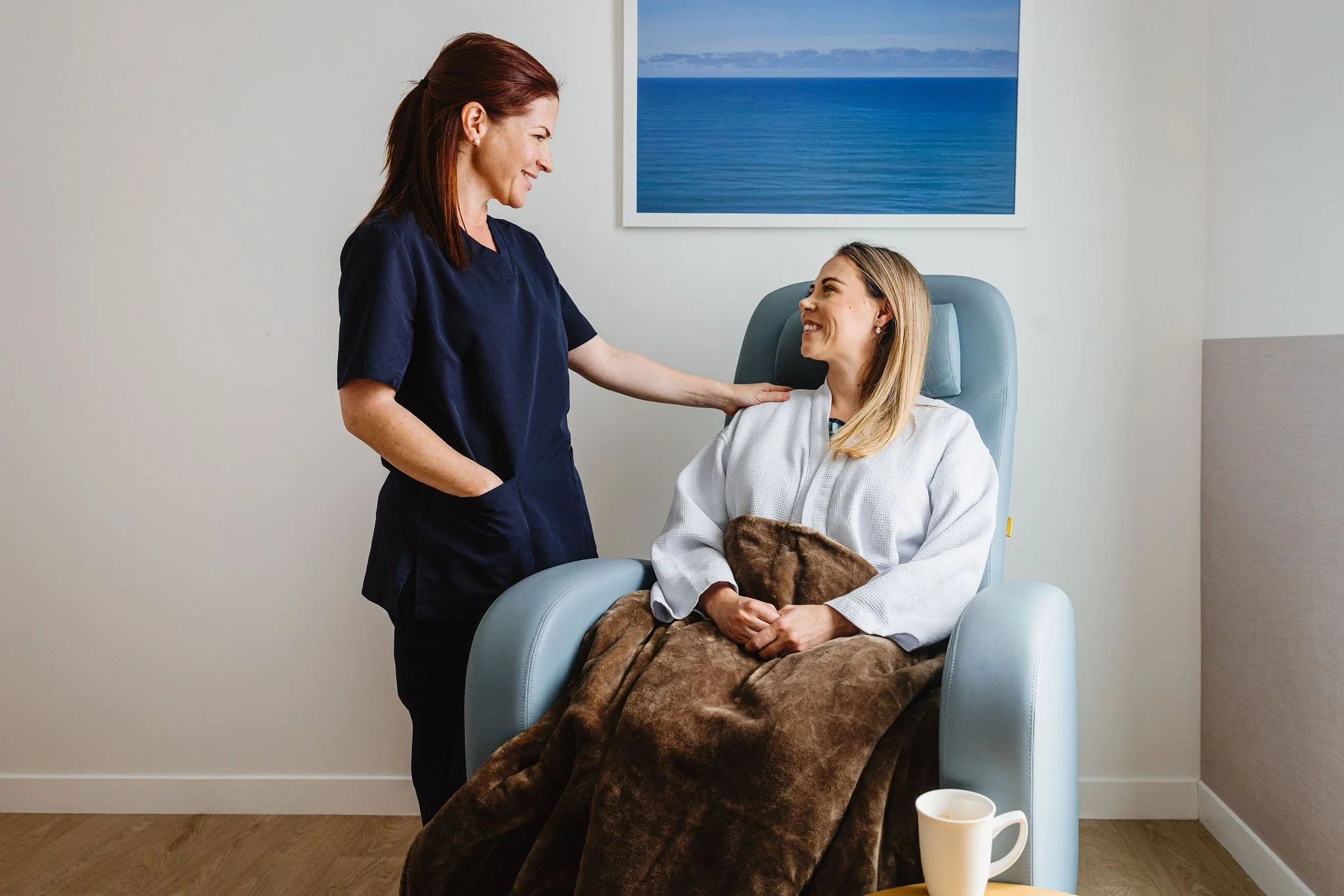
5. What this means when you come to see me
At Origin Aesthetics, every new patient consultation includes an assessment of your facial anatomy. For treatments in higher-risk areas — particularly around the nose, mid-face, and temples— ultrasound assessment is part of my usual approach where it is clinically useful.
This approach takes more time than a rapid injection appointment. I am comfortable with that. My aim is not to see the most patients in a day; it is to understand your face well enough to treat it well.
If you have had filler placed elsewhere and are considering treatment with me, ultrasound allows me to assess what is present before we proceed. That information often shapes what I recommend — and occasionally, it changes my recommendation entirely.
6. A final thought
There is a version of aesthetics medicine that is fast, transactional, and focused on throughput. I respect that many people are looking for that — quick, affordable, uncomplicated.
That is not what I offer.
What I offer is the approach of a specialist who has spent a career thinking carefully about anatomy, precision, and risk — and who brings that same thinking to every face she treats.
If that resonates with you, I would love to meet you.